 Diabetes In Control. A free weekly diabetes newsletter for Medical Professionals. News and information for Medical Professionals.
How Does Hyperglycemia Cause Complications?
Microvascular complications occur in cells and tissues which are unable to limit glucose transport in the face of hyperglycemia (particularly the retina, the mesangium in the kidney and Schwann cells). Several metabolic pathways and secondary messengers have been implicated in these tissues and the main ones are dealt with here.
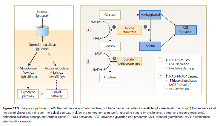
In this pathway, the rate-limiting enzyme, aldose reductase, reduces glucose to its sugar alcohol, sorbitol. This is then oxidized by sorbitol dehydrogenase into fructose. Aldose reductase is a ubiquitous enzyme found in many tissues but specifically nerve cells, retinal cells, the glomerulus and kidney tubule, and blood vessel walls. The pathway is normally inactive but in the presence of hyperglycemia there is increased flux leading to accumulation of intracellular glucose and glucose-derived substances, such as methyl glyoxal and acetol, which can rapidly glycate proteins (see below). Sorbitol does not diffuse easily across cell membranes and damage may occur because of osmotic stress (currently thought to be less likely although may be operative in the lens of the eye in the formation of cataract). Alternative mechanisms involve decreased levels of nicotinamide adenine dinucleotide phosphate hydrogen (NADPH), and increases in nicotinamide adenine dinucleotide plus hydrogen (NADH). The result of these changes is activation of protein kinase C and the promotion of advanced glycation endproduct formation (see below). Moreover, these changes result in increased oxidative stress (see below).
Advanced glycation endproducts (AGEs) are formed by the reaction of glucose and other glycating compounds, such as methylglyoxal, with proteins (an analagous process to the formation of glycated hemoglobin), and other long-lived molecules, such as nucleic acids. Early glycation products are reversible, but eventually they undergo irreversible change through cross-linking (Figure 14.10).
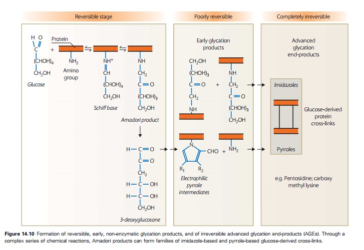
Advanced glycation endproducts can cause damage and ultimately complications of diabetes in two ways; firstly, as a result of cross-linkage of matrix proteins, such as collagen and laminin, leading to thickening and stiffening of blood vessels which can affect permeability and elasticity. Secondly, AGE-modified circulating proteins bind to specific receptors (RAGEs – three subtypes have now been described) on several types of cell, including monocyte/ macrophages, glomerular mesangial cells and endothelial cells. This binding leads to the generation of reactive oxygen species, activation of secondary messengers such as protein kinase C (PKC), release of transcription factor NF ê B and stimulation of cytokine and growth factor production, which can result in inflammatory cell adhesion (via increased VCAM-1), procoagulant protein expression and increased vascular permeability (via VEGF) (Figure 14.11).

Recently a circulating soluble RAGE has been identified which appears to mop up AGEs; reduced levels of this scavenger have been linked with increased atherosclerosis.
Several experimental agents that either reduce AGE formation or break cross-links have been tested in animals and humans. The first of these, aminoguanidine, has proven to be too toxic but others are in phase II trials.
Extrinsic AGEs are found in tobacco smoke and processed foods (notably roasted meats and some soft drinks). High levels of dietary AGEs have been associated with accelerated atherosclerosis in animals but their role in human disease is uncertain.
Protein kinase C is an enzyme that phosphorylates several target proteins. It exists in many isoforms and is activated by diacylglycerol which is a direct product of increased glucose flux and increased glycolysis. Overactivity of PKC has been implicated in increased vascular permeability and blood flow, particularly in the retina (Figure 14.12).
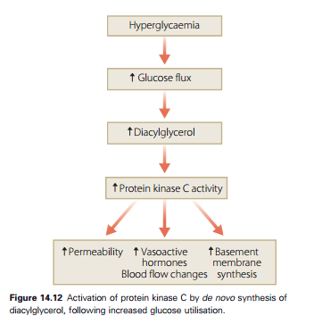
Interest in this pathway has been stimulated by the development of a PKC beta inhibitor, ruboxistaurin, which was shown in experimental animals to reduce the development of retinopathy. Trials in humans have shown benefit in advanced eye disease but confirmatory studies are awaited (see Chapter 15).