 Diabetes In Control. A free weekly diabetes newsletter for Medical Professionals. News and information for Medical Professionals.

Insulin is synthesized in and secreted from the β-cells within the islets of Langerhans in the pancreas. The normal pancreas has about 1 million islets, which constitute about 2-3% of the gland’s mass. All of the islet cell types are derived embryologically from endodermal outgrowths of the fetal gut. The islets can be identified easily with various histological stains, such as hematoxylin and eosin (Figure 5.1), with which the cells react less intensely than does the surrounding exocrine tissue. The islets vary in size from a few dozen to several thousands of cells and are scattered irregularly throughout the exocrine pancreas….

Islet cells interact with each other through direct contact and through their products (e.g. glucagon stimulates insulin secretion and somatostatin inhibits insulin and glucagon secretion) (Figure 5.3). The blood flow within the islets is organized centrifugally so that the different cell types are supplied in the sequence β → α → δ . Insulin also has an ‘autocrine’ (self-regulating) effect that alters the transcription of insulin and glucokinase genes in the β cell.
The pancreatic islets are densely innervated with autonomic and peptidergic nerve fibers (Figure 5.4). Parasympathetic innervation from the vagus stimulates insulin release, while adrenergic sympathetic nerves inhibit insulin and stimulate glucagon secretion. Other nerves that originate within the pancreas contain peptides such as vasoactive intestinal peptide (VIP), which stimulates the release of all islet hormones, and neuropeptide Y (NPY) which inhibits insulin secretion. The overall importance of these neuropetides in controlling islet cell secretion remains unclear.
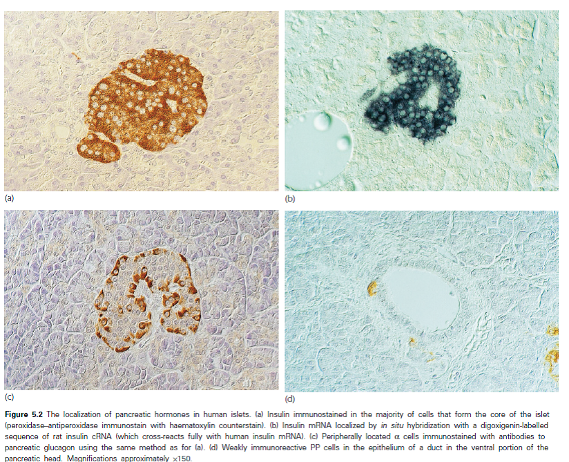

Pancreatic β cells may change in size, number and function during normal ageing and development (Figure 5.5). Β-cell mass is determined by the net effect of four independent mechanisms: (i) β cell replication (i.e. division of existing β-cells), (ii) β-cell size, (iii) β-cell neogenesis (i.e. emergence of new β-cells from pancreatic ductal epithelial cells) and (iv) β-cell apoptosis. The contribution made by each of these processes is variable and may change at different stages of life.
Insulin synthesis and polypeptide structure
The insulin molecule consists of two polypeptide chains, linked by disulphide bridges; the A-chain contains 21 amino acids and the B-chain 30 amino acids. Human insulin differs from pig insulin (an animal insulin which was used extensively for diabetes treatment prior to the 1990s) at only one amino acid position (B30) (Figure 5.6).


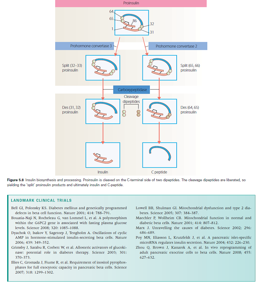
Insulin is synthesised in the β-cells from a single amino acid chain precursor molecule called proinsulin (Figure 5.8). Synthesis begins with the formation of an even larger precursor, preproinsulin, which is cleaved by protease activity to proinsulin. The gene for preproinsulin (and therefore the ‘gene for insulin’) is located on chromosome 11. Proinsulin is packaged into vesicles in the Golgi apparatus of the β-cell; in the maturing secretory granules that bud off it, proinsulin is converted by enzymes into insulin and connecting peptide (C-peptide).
Insulin and C-peptide are released from the β-cell when the granules are transported (‘translocated ‘) to the cell surface and fuse with the plasma membrane (exocytosis) (Figure 5.9). Microtubules, formed of polymerized tubulin, probably provide the mechanical framework for granule transport, and myosin and other motor proteins such as kinesin, may provide the motive force that propels the granules along the tubules. Although the actin cytoskeleton is a key mediator of biphasic insulin release, cyclic GTPases are involved in F-actin reorganization in the islet β cell and play a crucial role in stimulus-secretion coupling.

This ‘regulated pathway’, with almost complete cleavage of proinsulin to insulin, normally carries about 95% of the β-cell insulin production (Figure 5.10). In certain conditions, such as insulinoma and type 2 diabetes, an alternative ‘constitutive’ pathway operates, in which large amounts of unprocessed proinsulin and intermediate insulin precursors (‘split proinsulins’) are released directly from vesicles that originate in the endoplasmic reticulum.
Glucose is the main stimulator of insulin release from the β-cell, which occurs in a characteristic biphasic pattern-an acute ‘ first phase ‘ that lasts only a few minutes, followed by a sustained ‘ second phase ‘ (Figure 5.11). The first phase of release involves the plasma membrane fusion of a small, readily releasable pool of granules; these granules discharge their contents in response to both nutrient and non-nutrient secretagogues. In contrast, second-phase insulin secretion is evoked exclusively by nutrients. The shape of the glucose-insulin dose-response curve is determined primarily by the activity of glucokinase, which governs the rate-limiting step for glucose metabolism in the β-cell. Glucose levels below 5 mmol/L (90 mg/dL) do not affect insulin release; half-maximal stimulation occurs at about 8 mmol/L (144 mg/dL).
Glucose must be metabolized within the β-cell to stimulate insulin secretion (Figure 5.12). It enters the β-cell via the GLUT-2 transporter and is then phosphorylated by glucokinase, which acts as the ‘ glucose sensor ‘ that couples insulin secretion to the prevailing glucose level. Glycolysis and mitochondrial metabolism produce adenosine triphosphate (ATP), which closes ATP-sensitive potassium (KATP) channels. This in turn causes depolarization of the β-cell plasma membrane, which leads to an influx of extracellular calcium through voltage-gated channels in the membrane. The increase in cytosolic calcium triggers granule translocation and exocytosis. Sulphonylureas stimulate insulin secretion by binding to a component of the KATP channel (the sulphonylurea receptor, SUR-1) and closing it. The KATP channel is an octamer that consists of four K+-channel subunits (called Kir6.2) and four SUR-1 subunits.

There is a significant difference between the insulin secretory response to oral glucose compared with the response to IV glucose-a phenomenon known as the ‘ incretin effect ‘ (Figure 5.13). The incretin effect is mediated by gut-derived hormones, released in response to the ingestion of food, which augment glucose-stimulated insulin release. In particular, there are two incretin hormones: glucagon-like peptide-1 (GLP-1) and gastric inhibitory polypeptide (GIP). Both augment insulin secretion in a dose-dependent fashion.
GLP-1 is secreted by L cells and GIP is secreted by K cells in the wall of the upper jejunum.
In patients with type 2 diabetes, GLP-1 secretion is diminished (Figure 5.14). However, in contrast to GIP, GLP-1 retains most of its insulinotropic activity. GIP secretion is maintained in type 2 diabetes, but its effect on the β-cell is greatly reduced. GLP-1 also suppresses glucagon secretion from pancreatic α-cells, and has effects on satiety and gastric emptying. There is also considerable interest in the trophic effects of GLP-1 on β cells.
Insulin receptor signalling
Insulin exerts its main biological effects by binding to a cell surface receptor, a glycoprotein that consists of two extracellular α-subunits and two-subunits that span the cell membrane. The receptor has tyrosine kinase enzyme activity (residing in the β subunit), which is stimulated when insulin binds to the receptor. This enzyme phosphorylates tyrosine amino acid residues on various intracellular proteins, such as insulin receptor substrate (IRS)-1 and IRS-2, and the β subunit itself (Figure 5.15) (autophosphorylation). Tyrosine kinase activity is essential for insulin action.


Glucose is transported into cells by a family of specialized transporter proteins called glucose transporters (GLUTs) (Figure 5.17). The process of glucose uptake is energy independent. The best characterized GLUTs are:
- GLUT-1: ubiquitously expressed and probably mediates basal, non-insulin mediated glucose uptake
- GLUT-2: present in the islet β-cell, and also in the liver, intestine and kidney. Together with glucokinase, it forms the β cell’s glucose sensor and, because it has a high Km, allows glucose to enter the β-cell at a rate proportional to the extracellular glucose level
- GLUT-3: together with GLUT-1, involved in non-insulin mediated uptake of glucose into the brain
- GLUT-4: responsible for insulin-stimulated glucose uptake in muscle and adipose tissue, and thus the classic hypoglycemic action of insulin
- GLUT-8: important in blastocyst development
- GLUT-9 and 10: unclear functional significance.
Most of the other GLUTs are present at the cell surface, but in the basal state GLUT-4 is sequestered within vesicles in the cytoplasm. Insulin causes the vesicles to be translocated to the cell surface, where they fuse with the membrane and the inserted GLUT-4 unit functions as a pore that allows glucose entry into the cell. The process is reversible: when insulin levels fall, the plasma membrane GLUT-4 is removed by endocytosis and recycled back to intracellular vesicles for storage (Figure 5.18).


Next Week: Epidemiology and aetiology of type 1