 Diabetes In Control. A free weekly diabetes newsletter for Medical Professionals. News and information for Medical Professionals.
The dominance of ?-cell functional impairment
As discussed earlier, the available evidence indicates that an average 30% loss of β-cells is present in the islets of patients with T2DM. However, based on a series of considerations it is unlikely that this β-cell deficit alone is the cause of most cases of diabetes. First, in vivo assessment of β-cell function consistently shows a >50% reduction in patients with overt T2DM as compared to nondiabetic subjects, a difference that is amplified when using intravenous glucose [27,77,78] (Figure 24.3).

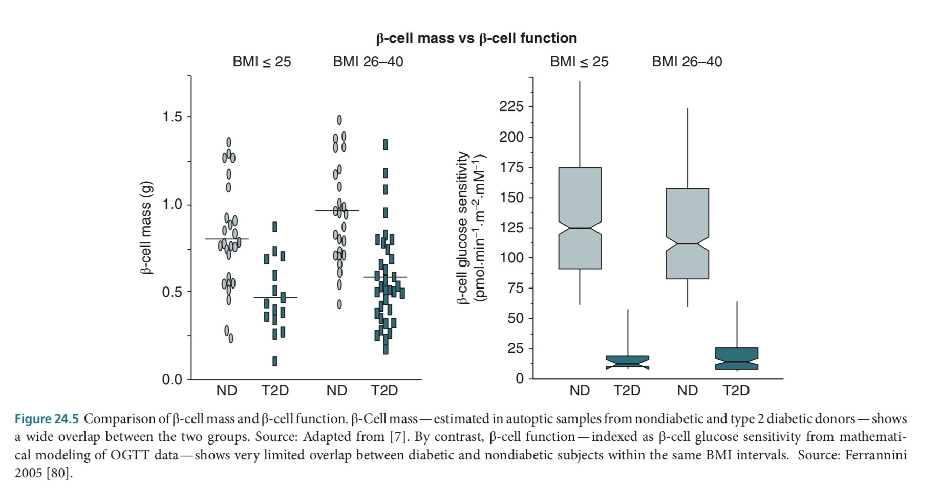
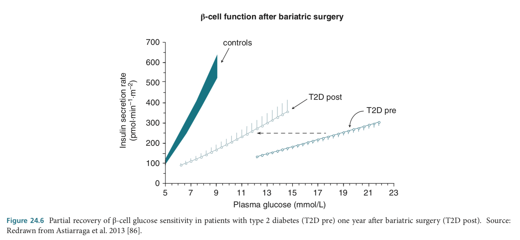
Thus, when β-cell function is indexed as β-cell glucose sensitivity (i.e., the slope of the insulin secretion/plasma glucose dose – response [79], type 2 diabetic patients show a marked inability to respond to changing glucose levels with a prompt rise in insulin release; this deficiency is accentuated in the presence of severe fasting hyperglycemia [80] (Figure 24.4). 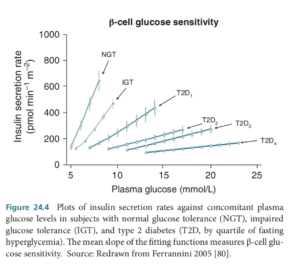 Of note, while β-cell mass estimates in type 2 diabetic patients overlap with those of nondiabetic subjects widely, the distribution of β-cell functional indices is well separated between type 2 patients and controls (Figure 24.5). It is therefore quantitatively incongruous that a deficit in β-cell mass can be the sole cause of the functional impairment. Secondly, in pancreatectomized patients and in subjects who undergo partial pancreas removal for the purpose of living organ donation, diabetes tends to develop more frequently over the years when β-cell mass declines by more than 50% [81–84]. Thirdly, bariatric surgery is followed by long-lasting resolution of diabetes in a high proportion of type 2 diabetic subjects, in some cases with full recovery of β-cell functional properties [85 – 87] (Figure 24.6). Finally, it has been clearly shown that any treatment resulting in amelioration of glycemic control is associated with partial reversal of β-cell dysfunction [88 – 90]. All this is in line with the findings of detailed morphometric studies, showing a very large variability in islet mass, with a wide overlap between nondiabetic and type 2 diabetic individuals [7] (Figure 24.1). Moreover, studies done in parallel with light and electron microscopy have shown that a proportion of β-cells might not be evidentiated by insulin immunostaining, due to low content of insulin granules, indicating the presence of living, but functionally incompetent β-cells [15]. As a consequence, β-cell secretory dysfunction is the dominant defect in the majority of diabetic patients.
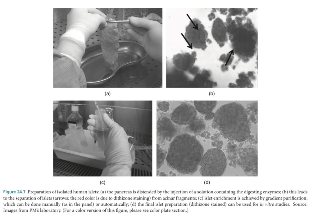
 This point can also be addressed by studying isolated human islets, which under certain circumstances can be prepared from the pancreas of organ donors (Figure 24.7) [91 – 93]. Although initial work was not able to show differences in insulin secretion
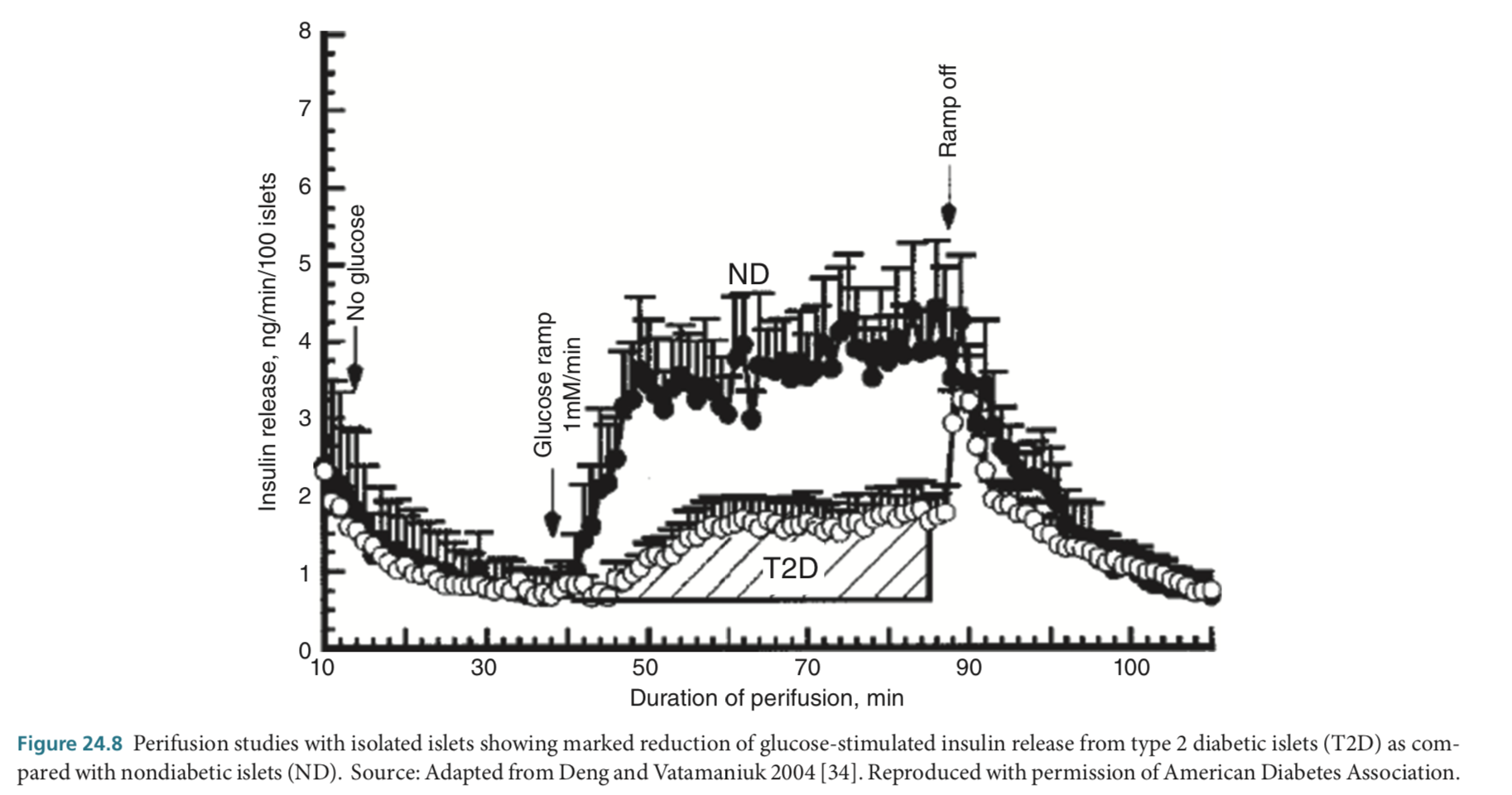
 between nondiabetic and type 2 diabetic islets [94,95], studies over the last 2 – 3 decades have consistently demonstrated defects of insulin release from type 2 human diabetes islets [18,36,96 – 98]. In a report published in 1994, it was shown that the release of insulin evoked by glucose was lower in type 2 diabetic than in nondiabetic islets [96]. However, the secretory response to a combination of L-leucine and L-glutamine appeared less severely altered [96]. In a more recent study, islets isolated from eight diabetic and nine nondiabetic donors were evaluated by in vitro islet perifusion experiments [98]. Basal insulin secretion was similar between normal and diabetic islets. However, the islets from diabetic donors released less total insulin in response to increasing glucose levels and also exhibited a higher threshold for the initiation of insulin secretion (Figure 24.8). In addition, an equivalent amount of type 2 diabetic islets did not fully correct hyperglycemia when transplanted into diabetic mice in comparison with normal islets [98]. In another study, when insulin secretion was measured in response to glucose, glibenclamide, and arginine in isolated nondiabetic and type 2 diabetic islets, again no significant difference in insulin release in response to 3.3 mmol L−1 glucose was observed [36]. However, when challenged with 16.7 mmol L−1 glucose, diabetic islets secreted significantly less insulin than nondiabetic islets. Insulin secretion during glibenclamide and arginine stimulation was also lower from diabetic islets than from control islets; of note, in this series type 2 diabetic islets released more insulin in response to non-glucose stimuli than in response to glucose. Interestingly, similar changes in insulin secretion can be induced in nondiabetic human islets by prolonged exposure to increased levels of free fatty acids (in particular, palmitate) or glucose [31,99 – 102], mimicking findings reported in in vivo studies [103,104]. All these data clearly indicate that type 2 diabetic β-cells have defective insulin secretory behavior, which contributes to the development of hyperglycemia at least in part independently of β-cell mass.
Disruption of ?-cell dialogue in the islet
It has long been recognized that β-cells dialogue with each other and with the other islet endocrine cells, mainly through autocrine and paracrine communication [105 – 111]. This is made possible by the peculiar organization of endocrine cells in human islets [3,112] — allowing more than 70% of β-cells to be in contact with non-β-cells [112] — and the abundance of gap junctions (containing connexin proteins) and other cell – cell adhesion complexes [109,113 – 115]. In addition, endocrine cells could influence one another by the action of the respective hormones via intra-islet blood flow [116,117]. Finally, nonhormonal endocrine cell products (such as ATP and zinc) and neurotransmitters (released either by islet cells or by sympathetic and parasympathetic nerve fibers) influence β-cell function [109 – 111]. Disruption of these multiple levels of β-cell dialogue in human T2DM has been hypothesized. A well-known feature of T2DM is hyperglucagonemia [118–121], but studies on the relative role of α-cell number and function have given conflicting results. Deng et al. [34] found that the proportion of glucagon-containing cells was significantly increased (52 vs. 35%) in their 14 type 2 diabetic as compared to 14 nondiabetic organ donor pancreases, whereas ex vivo glucagon release in isolated islet perifusion studies did not differ. However, in a larger series of 50 type 2 diabetic and 52 nondiabetic autototic samples, it was shown that α-cells mass was essentially the same in type 2 diabetic (366mg) and nondiabetic subjects (342 mg), and was not influenced by sex, BMI, or type of diabetes treatment [122]. Likewise, no difference in the number of α cells between type 2 diabetic and nondiabetic cases was shown by electron microscopy evaluation of pancreatic specimens from organ donors [15]. Nevertheless, due to the concomitant reduction of β-cells, in these studies the ratio of α cells to β-cells was significantly higher in diabetic islets [34,122]. This suggests that, on the one hand the reduction of intra-islet insulin in T2DM may contribute to a disproportionately elevated glucagon secretion [121], which could be exacerbated by defective glucose regulation of glucagon secretion (due to impaired metabolic control of the α cell ATP-regulated K+ (KATP) channels [123]); on the other hand, exposure to increased intra-islet glucagon concentrations could have adverse effects on the β-cell [124].
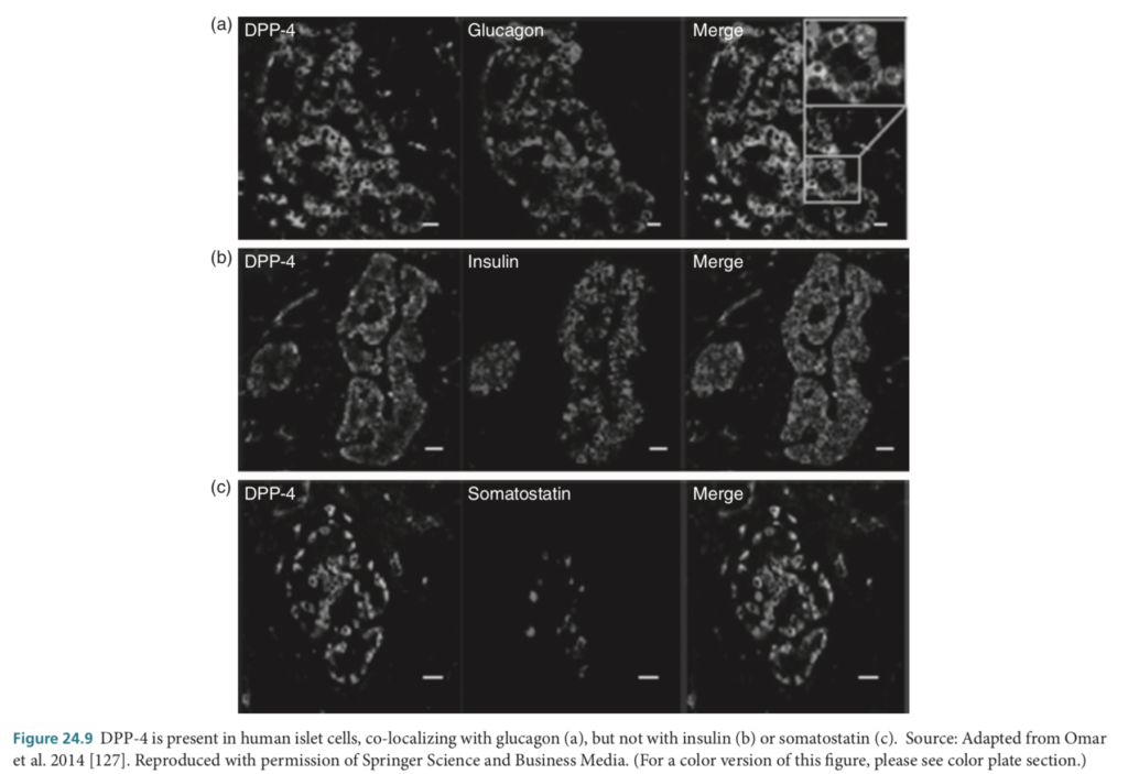 Other features of α cells could influence the β-cell in human T2DM. In fact, subpopulations of α cells have been shown to be able to process preproglucagon by proconvertase 1/3 to produce glucagon-like peptide-1 (GLP-1) [125,126]. In addition, ex vivo active GLP-1 release is higher in islets from type 2 diabetic individuals [126], and recent data show the presence of dipeptidylpeptidase-4 (DPP-4, the GLP-1 inactivating enzyme) in human α cells (Figure 24.9), with reduced DPP-4 activity in type 2 diabetic islets [127]. These findings are in line with the concept that, under given circumstances human α cells could modulate their phenotype to produce GLP-1 in an attempt to protect the endangered β-cells [125–129]; this phenomenon could include α-cell production of glucose-dependent insulinotropic polypeptide, GIP [130]. This novel information is relevant to the controversial issue of the role of incretin-based therapy in T2DM [131 – 135].
Recent observations have pointed to yet another abnormality of T2DM islets, namely, altered endocrine cell arrangement. Especially in larger islets, architecture is sparse, and spatial cell organization appears more dispersed [10,15]. Given the importance of the above described cell-to-cell communication for normal islet cell function [105 – 115], this morphologic feature may have physiologic impact. Interestingly, work has shown that connexin-36 (Cx36, a key component of gap junctions) correlates with insulin gene expression in both nondiabetic and type 2 diabetic islets [136]. In addition, exposure of human islets to lipotoxic conditions reduces Cx36 expression, disrupts islet cell architecture, and decreases glucose-stimulated insulin secretion [137].