 Diabetes In Control. A free weekly diabetes newsletter for Medical Professionals. News and information for Medical Professionals.
 Pancreatic anatomy and morphology
The pancreas is located in the upper abdominal cavity, in close proximity to both the duodenum and spleen (Figure 5.1(a)). In humans, pancreatic weight ranges from 40–150 g [1,2]. Embryonically, the pancreas develops from two separate buds of the primitive foregut, yielding duodenal (ventral) and splenic (dorsal) lobes [3]. Once developed, these constitute the head and body/tail, respectively.These regions of the pancreas are similar, aside from some differences in the distribution and composition of pancreatic islets (discussed later).
Functionally, the pancreas comprises two independent compartments, the exocrine and endocrine pancreas, which derive from common endodermal precursor cells during development [3]. The exocrine pancreas accounts for the vast majority of pancreatic mass and consists of lobules, each comprising acini that connect into a network of ducts (Figure 5.1(b)).
Acinar cells demonstrate a characteristic morphology at the electron microscopy level including electron-dense zymogen granules and an extensive endoplasmic reticulum (ER) network. This ultrastructural organization is consistent with the chief function of the exocrine pancreas, namely to secrete digestive enzymes including amylase and trypsin via the pancreatic ductal system into the gut. The endocrine pancreas comprises islets of Langerhans, roughly spherical structures that contain hormone-producing cells. Islets constitute only a small minority of pancreatic mass but are critically important for metabolism throughout the body, and especially for maintenance of glucose homeostasis. This was first shown in 1890 by studies in which pancreatectomy in dogs resulted in diabetes [4]. In the human pancreas, it has been estimated that there are one to two million islets scattered throughout the exocrine pancreas, which together comprise approximately 2% of pancreatic mass [5,6]. While the function of these two pancreatic compartments differ significantly, they exist in close proximity to one another, and evidence exists for interactions between them. Specifically, islets are distributed throughout the exocrine pancreas. Islet hormone-rich blood perfuses the exocrine pancreas [7], and the islet hormones, insulin and pancreatic polypeptide, have been shown to stimulate amylase secretion from the exocrine pancreas, while glucagon inhibits amylase secretion [8–10]. This suggests that the functional status and viability of the islet can influence function/viability of the exocrine pancreas and potentially vice versa.

Islet composition and morphology
Despite the relatively small size of pancreatic islets, each comprising approximately 2000–4000 cells [11], every one is a complex mini-organ, containing numerous cell types (Figure 5.1(c)). Endocrine cells are, not surprisingly, the most abundant islet cell type and are characterized at the ultrastructural level by the presence of numerous secretory granules that contain endocrine hormones packaged and stored ready for immediate release in response to the appropriate stimulus. Additionally, endocrine cells contain a high density of mitochondria and abundant ER (although not so dense as the surrounding acinar cells).These endocrine cells are subcategorized based on their predominant hormone constituent, as follows.
Insulin-producing β cells are the predominant islet cell type. They were first identified in 1907 by silver staining [12] and were the second islet endocrine cell type to be described. β Cells comprise 50–80% of islet volume, depending on the species in question; rodent islets typically have a higher proportion of β cells (around 60–80%) [13,14], while nonhuman primate and human islets have a lower relative proportion (around 50–60%) [13–19] (Figure 5.2). The distribution of β cells within islets also differs among species. In rodents β cells are predominantly located in the core of the islet [13,14,19], while in nonhuman primate and human islets they are more evenly distributed throughout the islet (Figure 5.1(c)) [13,14,18]. Regardless of their location within the islet, for all species β-cell secretory granules have a characteristic appearance at the electron microscope level, with an electron-dense core that arises due to the formation of insulin hexamers which are cross-linked with zinc [11]. This granule core is surrounded by an electron-lucent “halo” [11]. Release of insulin fromβ cells is critically important in regulating blood glucose levels, predominantly acting to suppress hepatic glucose production and to enhance glucose uptake in insulin-sensitive tissues such as skeletal muscle and adipose tissue. Mechanisms of insulin release are discussed in detail in Chapters 7, 8, and 9. In addition to insulin, β cells also produce islet amyloid polypeptide (IAPP), which is co-localized with insulin in secretory granules and is therefore co-secreted with insulin [20].
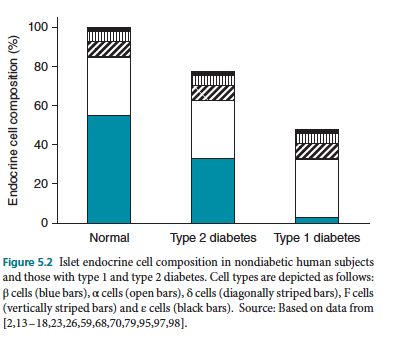 Glucagon-producing α cells were actually the first islet endocrine cell type to be described and are so called due to the alcohol fixation (“A”-cells) that was used to identify them [12]. These form the second most abundant endocrine cell type in the islet, but are far less abundant than β cells. In rodent islets, they account for around 15% of islet area [19], whereas in humans they are more abundant (around 35%; [13]) (Figure 5.2). Also, like β cells, α cells differ in their distribution within islets across species. In rodents, they are located almost exclusively in the mantle of the islet [13,14,19], while in human and nonhuman primate islets they are more evenly distributed throughout the islet (Figure 5.1(c)) [13,14,18]. Unlike β cells, whose abundance in islets is relatively constant throughout the pancreas, islets in the head of the pancreas are richer in α cells than those in the tail [19]. Glucagon-containing secretory granules differ in their appearance compared to insulin-containing granules, in that they lack an electron lucent halo. Thus, the entire secretory granule in the α cell is electron dense [21]. The hormonal effects of glucagon act to oppose those of insulin, stimulating hepatic glucose production, and being a critical mediator of the restoration of normal glucose levels following hypoglycemia. Glucagon secretion and its effects are described in Chapter 10.
Somatostatin-producing δ cells comprise the third islet endocrine cell type, being lower in abundance (<10% of islet cells) than either β or α cells (Figure 5.2). Their distribution within islets is similar to that of α cells, namely that in rodent islets δ cells occur predominantly in the mantle of the islet, while in primates and humans they are more evenly distributed throughout [13,14,18]. While few studies have systematically assessed δ-cell distribution in islets throughout the pancreas, δ-cell number does not appear to be markedly altered in islets from different regions of the pancreas [19]. Ultrastructurally, somatostatin-containing granules are homogeneous in appearance, similar to α-cell granules, but slightly less dense. The effects of islet-derived somatostatin are thought to be largely paracrine in nature due to its short half-life [22]. Somatostatin acts on receptors present on β and α cells, inhibiting both insulin and glucagon release.
Pancreatic polypeptide-producing F cells are less abundant still than δ cells, contributing on average less than 5% of islet cells. Again like α cells, their distribution differs throughout the pancreas, with islets in the head of the pancreas containing most of the pancreatic polypeptide-producing F cells [19,23]. Thus, islets in this region of the pancreas can contain up to 20% pancreatic polypeptide-containing cells, at the expense of α cells, which are decreased in this region. Conversely, in the rest of the pancreas, F cells only account for a 1–3% of islet cells [19,23] (Figure 5.2). F-cell granules are small, but similar in appearance to β-cell granules, containing an electron-dense core and electron-lucent halo. The release of pancreatic polypeptide from islets serves as an indicator of vagal outflow [24]. However, the function of pancreatic polypeptide, once released, is unclear. Some studies have suggested a role in inhibition of pancreatic enzyme and bile secretion, while others have suggested a role in modulation of food intake and energy expenditure [9,25].
Ghrelin-producing ε cells constitute the final known population of islet endocrine cells. These were first described during embryonic development [26], and were originally thought to reflect a population of multi-hormone positive cells. However, they have been shown to be present in adult islets, and are now considered a bona fide islet cell type [26]. Several studies have shown that ghrelin can suppress insulin release in humans and rodents [27,28]. However, whether this occurs due to a paracrine effect of islet-derived ghrelin or due to circulating ghrelin, which is largely produced in the stomach, is unclear at this time.
Click here to view all Chapter 5 references.